Veterinary Virology and Mother Nature’s Kitchen

By Claire Birkenheuer, PhD

Post-doctoral researcher Dr. Joel Baines Lab, Pathobiological Sciences Department, Louisiana State University


I got my PhD in microbiology studying a little known retrovirus that infects walleye fish, called walleye dermal sarcoma virus (WDSV).  I did my research in the Quackenbush/Rovnak lab at Colorado State University.  When I started my PhD, I really wanted to study a human virus that caused serious disease, yet somehow I ended up at a vet school in a lab that studied a virus that caused a benign disease in a fish that I only knew of because my grandfather went fishing.  This experience turned out to be the best PhD project, and it really changed my way of thinking about medicine, basic research, and how the veterinary field contributes to modern human medicine.

For my project, I was assigned to study the molecular mechanism behind retroviral cyclin, which is one of the three accessory proteins encoded by WDSV.  WDSV is an interesting virus because it is the only virus we know of that purposefully causes a cancer for its replication.  The virus infects fish at spawning in the spring.  The virus then activates cellular cancer genes in the skin which cause dermal sarcomas to grow.  These appear on the fish in the fall following the spring infection.

walleye-dsv-image

Walleye dermal sarcoma virus causes tumors to form on the skin of walleye fish (adapted from Rovnak and Quackenbush, 2010 Viruses)

The tumors grow throughout the winter.  During this time, the virus is not replicating, and the only viral transcripts produced are retroviral cyclin, and the Orf B protein.  When spawning is approaching the following year, the virus switches its expression profile, and starts replicating in the tumors it has grown.  This replication involves expression of the third accessory protein, Orf C, which causes the tumor cells to undergo apoptosis precisely at the time of spawning.  This event correlates with the tumor shrinking and falling off the fish, releasing virus particles in the water when all the fish are together mating.

You might be thinking Wow! What an interesting life cycle, but what does this have to do with human medicine.  However, when you think about it, there are many.  For example:

  • How do two viral proteins cause a dermal sarcoma?
  • What can this tell us about human skin cancers?
  • What is it about the Orf C protein that causes the tumors to fall off?
  • Could this OrfC protein be used as a therapeutic agent for human cancers that could be specifically delivered to cancer cells in a viral vector?
  • What does this tell us about pathogen-host evolution?

By being guided through these questions as a PhD student I gained an increased understanding of the importance of veterinary medicine to human medicine, as well as an awe for Mother Nature’s kitchen.  Could the cure for cancer be out there in nature?  If so, we really need to have scientists who explore these questions in veterinary medicine.  If anything, as scientists we must explore every possibility, and keep in mind that Mother Nature has many tricks up her sleeve.  Whether it is in snake, or spider venom, a little-known fish virus, or some plant extract, Mother Nature may have already come up with an answer to some of medicines most needed problems.  It is important to fund and include veterinary science, and basic biology in our quest for cures to some of the most devastating human diseases.  Although I did not study a human pathogen, WDSV taught me many valuable lessons.

Now, as a postdoc, I study Herpes Simplex Virus 1, and although it is a human pathogen which can cause serious disease, I fondly remember and somewhat miss that fish retrovirus and all the possibilities it had to offer.  Perhaps someday I will get a chance to go back to it.  I can only hope that funding agencies see the value and human benefit in supporting research projects like the one I took part in with WDSV.

Judging a bacterium by its cover: immune cell–symbiont communication in the Hawaiian Bobtail squid/Vibrio fischeri symbiosis

mcanulty1By Sarah McAnulty (@SarahMackAttack), a doctoral candidate in the Nyholm lab (Department of Molecular and Cell Biology, University of Connecticut)


The first thing everyone tells me when they hear about my work is, “Your squid are so cute!” and guys. They’re right. I mean look at them:

It doesn’t get much more adorable than that, and I’m the first to admit it. But their cute-factor isn’t what gets me jazzed about the Hawaiian bobtail squid.What excites me is what these little squid have going on under the hood. Let me back up for a second and tell you why we study these squid in the first place.

Hawaiian bobtail squid are nocturnal predators on the reef, but much like many other squid, they are effectively swimming protein bars that bigger fish can’t wait to get their jaws on. Being in this position, many cephalopods have become extremely effective at hiding. Many of you may have already seen this video of octopus, but it’s a great demonstration of how well these animals do at hiding from the gaze of predators.


Octopus and cuttlefish have both become world class camouflagers, and Hawaiian bobtail squid have too, but instead of using color changing skin cells (chromatophores) to hide, the squid has developed a symbiosis with a bioluminescent bacterium called Vibrio fischeri. The squid keep these bacteria in a specialized “light organ” on their underside and are able to control the amount of light that escapes from the organ to match the moonlight coming from above.

Here’s a video of a closely related species of bacteria, Photobacterium leiognathi:

When squid first hatch, they need to find this bioluminescent bacterium swimming in the seawater. They’re extremely efficient at finding their partner, but what’s even more extraordinary to me, is that their immune cells can tell the difference between V. fischeri and other types of bacteria. This is totally nuts because compared to the human immune system, the squid immune system isn’t super complicated. In fact as far as we know, squid don’t have any kind of adaptive immunity or immunological memory at all. What we’ve noticed studying these immune cells (called hemocytes) is that not only do they treat V. fischeri differently, but the V. fischeri needs to be present in the light organ in order to maintain this tolerant hemocyte behavior. Previous work used antibiotics to remove V. fischeri from the squid, and after the treatment, squid immune cells no longer tolerated V. fischeri. So it seems that the bacterium is communicating, “Don’t eat me!”, to the immune system constantly.

The goal of my work is to understand how V. fischeri communicates with hemocytes to identify itself, and what hemocytes change about themselves and their behavior to accommodate for living in a symbiosis with V. fischeri. I’m exploring this by watching immune cells and how they interact with bacteria when I either raise squid with or without Vibrio fischeri. I’ve also compared how they treat various mutants of V. fischeri in an attempt to figure out what it is on the surface of V. fischeri or released from V. fischeri that’s the identifying factor. To do this, I bleed squid and then isolate the hemocytes from their blood (don’t worry, they survive the quick blood draw and are totally anesthetized when I draw blood from them).

mcanulty4

Squid blood is blue!

I then label hemocytes with a live-cell stain called Cell Tracker Deep Red. This allows me to see my hemocytes (pink), and various species of bacteria (blue and yellow) all at the same time. I then visualize them on a confocal microscope. Confocal microscopes are particularly cool because they can focus on just one thin section of whatever you’re looking at, and then take pictures through multiple thin seconds. You end up with 3-D images, and for my work I end up taking 3D videos. Having beautiful videos is a definite bonus for working with this system.

This is what my data end up looking like! Hemocyte is pink, and two bacterial species are in blue and yellow.

In addition to my microscopy, our lab is also working on proteomics with squid immune cells. We’re hoping that by identifying the proteins that change between squid that are raised with and without V. fischeri, we’ll be able to determine what affects this change in hemocyte behavior after the addition of V. fischeri.

Squid are cute, hemocytes are cool, but is this important? (Spoiler: totally)

You might be asking yourself why any of this is important. Ok, your squid is cute, but why do we care what its immune cells are doing?  Well, the thing about the human immune system, is that beneath all the bells and whistles of the adaptive immune system (antibodies, etc), we still have an innate immune system, which the squid has too. Partnering with bacteria is an ancient and widespread phenomenon in animals. Everything from the lowly hydra to a giraffe lives in association with bacteria (there are even efforts underway to characterize the microbiomes of all zoo animals which is pretty fun).  If we learn how this squid can tell the difference between “good” and “bad” bacteria, we’re going to learn something about ourselves too. A lot of really nasty stuff happens when your gut microbiome gets out of whack, as you’ve probably heard. Messed up microbes are associated with many diseases and disorders, including diabetes, obesity, autism and MS. It’s really essential for us to understand how our immune systems are sensing who’s there, in order to see how these associations go wrong. We use the squid because the symbiosis with V. fischeri is super convenient, since the light organ only houses V. fischeri. Compare that to a mouse that can house up to 1000 species of bacteria. That’s a lot of noise to work though, so having one bacterial partner, especially one that you can raise squid without if you want to, makes for a great animal model.mcanulty6

If you want to learn more, there’s plenty of information out there about squid and this symbiosis.

For further reading on hemocytes and the squid-vibrio symbiosis: http://bit.ly/2hY1ZqD

For more on squid biology and grad life/career advice, follow me on tumblr squidscientistas.tumblr.com

Follow me on twitter @SarahMackAttack

Come out, come out, wherever you are: searching for rare frogs in a population devastated by an emerging infectious disease

By Graziella DiRenzo, PhD, Post-doctoral research associate in Dr. Elise Zipkin’s lab in the Department of Integrative Biology at Michigan State University

For more info of the Zipkin Quantitative Ecology lab, watch the new lab video:


Tropical cloud forest in the mountains of El Copé, Panama.− Night falls. It’s time. I scour the dense forest and streams, searching for tiny green and brown frogs camouflaged against green vegetation or brown leaf litter. I keep moving although the fear of encountering a venomous snake or a jaguar the next place my headlamp illuminates chills my bones. No genius needs to tell me that I’m not finding all the frogs in the forest. Who can blame me? They are really hard to see!

This continued for months. Night after night…

direnzo-fig1

Nicole Angeli and I doing a night survey in El Cope, Panama

Temperate office in the lowlands of College Park, Maryland.− A strand of hair tickles down my arm, and I quickly swat at it. To my surprise it isn’t a tropical mosquito or a spider that I picked up while brushing against a palm tree, it was merely a single hair from my head− not the immediate threat my body registered.

After my first field season in El Copé, Panama, I immediately started to worry about the possibility that I couldn’t finish my dissertation because I couldn’t find enough frogs.

Why was I so determined to write a dissertation on frogs that were really hard to find?

Because a fungal pathogen named Batrachochytrium dendrobatidis, typically referred to as Bd, spread through Central America in the early 2000s, causing the extinction and decline of tens of hundreds of amphibian species.

Because amphibians are the only known host of Bd, one of the only types of fungus that kills vertebrates.

And, because not all amphibians respond the same to Bd infection.

My project focused on understanding how amphibians persist with Bd, and how Bd transmission works in this system.

But, how in the world was I going to answer these questions when I couldn’t even find that many frogs?!?!

Good question. I definitely wasn’t the first person to consider how not detecting individuals that were present in a location during a search could affect the conclusions that we make.

I found myself in the realm of statistical models that takes advantage of count data collected from repeatedly searching the same area to calculate a detection probability (i.e., the probability an individual is found during a search, given that the individual is present)− known as “Royle models” or “Dail-Madsen models”. This was the very tool I needed in my statistical toolkit to outsmart those small, nearly invisible little suckers. The book that quickly became my sidekick, my go-to, my-everything was: Bayesian population analysis using WINBUGS by Marc Kéry and Michael Schaub. I recommend everyone have a copy handy for their statistical needs.


direnzo-fig2

Sachatamia albomaculata– a species of glass frog- mating. The male holds onto the female until she is ready to lay her eggs, and he will fertilize them.

The more I learned, the more I hungered for other statistical tools, and the more tools I acquired, the more ecologically-motivated questions I asked. The more I asked, the more I learned. Bringing me back to the beginning. This self-engulfing loop ran on repeat for the next 5 years of my PhD at the University of Maryland with Dr. Karen Lips. Check out the Lips’ lab video here:


But, my big problem now was that these models did not allow uninfected and infected individuals to have different survival, detection, and recruitment rates. Although, around this same time, scientists were beginning to add R code as appendices to their articles, this wasn’t enough for me to find an answer.

In the end, I developed a novel framework to estimate survival, detection, recruitment, and disease transition rates using count data of frogs. I am currently wrapping up this project, so stayed tuned for the published paper and the cool results!

Currently, in an effort to help others that are plagued by similar detection problems, I am:

(1) providing the code and materials I generate from research projects at: http://grazielladirenzo.weebly.com/code.html

And although your Bayesian or JAGS struggles are likely not exactly the same as mine, just know that I get a bad taste in my mouth when I see the words: Node inconsistent with parent values.

(2) blogging about Bayesian methods, commenting on the things I wish I knew before starting a project. Check it out if you ever have Bayesian problems: http://grazielladirenzo.weebly.com/bayes-the-way

(3) creating short videos that help understand the types of projects that different labs work on and short videos for undergraduate and teacher projects I co-advise (Check out Lips Lab REU Scholar or NSF RET project). All of these videos can be found on my YouTube channel: graziella direnzo:                                                                               https://www.youtube.com/channel/UCI9Ia_dsfzAAtJASS7cmqyg

I know this is one small drop in the ocean of science, but if more and more scientists share their simulation code and statistical tools, then we as a community can make bigger leaps in advancing our knowledge.

Sniffing out crime: vapor analysis for arson investigation

By Megan Harries, doctoral candidate in the Department of Chemistry and Biochemistry, University of Colorado-Boulder

I know it’s going to be a good day in the lab when “start a fire” is on my to-do list. As a PhD candidate working at the National Institute of Standards and Technology (NIST), on a fire-starting day I’m studying the vapor signature of an accelerant after it’s been burned in an arson fire.

Arson is a tricky crime. According to the National Fire Protection Association, only 21.8% of known cases are cleared and it’s likely that, because of the difficulty discerning arson from accident, that’s an overestimate. It’s one of the easiest crimes to get away with. Why? Part of the problem is with the forensic science. It may surprise you that a lot of evidence is left behind even after a fire has completely consumed a structure. There’s likely residue of accelerant remaining, if one was used, but its detection and identification requires very sensitive, robust sampling and analysis. Currently accepted methods don’t always get the job done.


Here’s an example of fire debris generation. The wood block has been soaked in WD-40 and, if you look closely as it burns, you can see boiling happen at the surface. The accelerant is evaporating, and the vapor is what really burns.

At NIST, I’m working to optimize a new method and device (called PLOT-cryo) for the collection of vapor samples like those important to arson fire investigation. I start controlled fires in the lab using an accelerant—we’ve studied diesel fuel, gasoline, kerosene, industrial solvents, etc.—as a means of generating samples of what we call arson fire debris. These samples become tests for my method of headspace analysis, the chemical characterization of the vapor that forms above and around a solid or liquid sample of interest. I enclose the fire debris in a metal paint can to develop and contain this headspace. After a certain period of time, I use PLOT-cryo to collect the headspace onto an adsorbent trap that captures its chemical signature. Injecting this headspace sample into an analytical instrument like a mass spectrometer allows me to discern its composition, and therefore, the chemical makeup of the fire debris itself.


picture1

This is the portable PLOT-cryo device, which was developed in our lab. At left, the sampling probe is inserted into a can containing arson fire debris. A vacuum generator inside the unit (right) provides the suction we use to pull vapor up through the probe and into the adsorbent trap, housed inside the plastic box.

Most commonly used accelerants are complex mixtures, which works in our favor as forensic scientists. A GCMS analysis of a mixture is like a fingerprint. By identifying its components and their quantities relative to one another, we can actually determine the identity of the fluid that was used. Because fuels like gasoline and diesel fuel vary regionally and across brands, it’s reasonable that we could even identify the brand name or the location of the gas station where it was purchased, if we had a sample or library to use as a reference.

figure_2_mh

A chromatographic analysis of a fire debris sample reveals the components of the accelerant used to start the fire (in this case, aviation gasoline). This chemical “fingerprint” of the accelerant residue is useful to investigators.

In addition to the arson work, we’ve tested the device in simulated forensic situations like food spoilage and safety, explosives detection, and to analyze the chemical composition of gravesoil.

This project has given me opportunities to work at the intersection of basic research, real world application, business, and communication to stakeholders. Beyond the technical work, I’m learning about the public dissemination of new research and how to talk with folks who are potential end users of a PLOT-cryo device. Our arson research has drawn the attention of an organization dedicated to educating judges about the latest forensic science techniques, and a group of them will visit NIST early next year for a two-day workshop about PLOT-cryo. One of my colleagues left NIST just a couple of months ago to start a business manufacturing the device and marketing it to vintners interested in monitoring the quality of their wines. I’m excited to see his progress as he gives the technology commercial life. I’ve benefited professionally and personally from seeing the whole life cycle of science happen here, from basic thermodynamic measurements of phase equilibrium, to instrument and method development, to optimization, to commercialization.

Take a moment to appreciate the role of vapor characterization in modern life. The Breathalyzer keeps millions of drivers safer. Working dogs locate illicit drugs, explosive devices, and disaster victims. Your own nose and brain characterize vapor so fluidly every day that you take for granted the smell of a good meal or a gas leak. Aroma and flavor are both mediated by interactions between vapor molecules and your nose’s receptors. If you remember just one thing from reading this post, I hope it’s this: vapor chemistry is important, and life would be pretty boring without it!

A day in the life of a hospital neuroscientist

By Jonathan Jackson, PhD

I am a cognitive neuroscientist on faculty in the Neurology department at Massachusetts General Hospital (MGH). I research the early detection of Alzheimer’s disease in a human research laboratory in Boston, Massachusetts. Working in a hospital may sound wildly different from running a lab in a university, but it is actually a lot more similar than you might think. If you’re considering a research career, please read on; this may be an attractive option that you may not have considered.

In many ways I am a traditional academic. I’m on the tenure track at Harvard Medical School (HMS), for example. I am required to generate, communicate, and evaluate research through peer-reviewed publications. We hold weekly lab meetings to discuss (read: argue about) the latest research findings from our own and other Alzheimer’s labs. There are dozens of committees at MGH that I may join in order to fulfill service obligations. I work with brilliant colleagues on complex problems. You won’t find patient beds on our floor; instead, we work in a mix of traditional cubicles and offices that you’d find at any university. Overall, the environment I work in, and career goals I strive toward wouldn’t feel out of place in any university setting.

lab-meeting

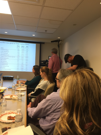
Like many labs, we have regular meetings to discuss the latest findings from our research. However, sometimes our meetings are attended by a film crew hoping to learn about breakthroughs in the fight against Alzheimer’s disease!

However, there are also a number of striking differences that might make working at HMS / MGH more (or less!) appealing for those looking to develop research careers outside of the typical college or university campus.

The biggest difference is in how my laboratory is structured. First of all, there aren’t any graduate students (or medical students)! Instead, we have more than 20 doctoral-level (PhDs, MDs, and PsyDs) individuals and 15 (mostly post-baccalaureate) research assistants performing full-time research around a large NIH-funded program project grant and several satellite grants. Collectively, we run the Harvard Aging Brain Study, a large prospective cohort of 400 older adults. Although many of us are on the tenure track, have obtained our own NIH or foundation funding, and have largely independent research careers, more senior lab members will mentor the younger ones, and tend to have more influence in how the overarching study is tweaked and conducted. Physicians and clinicians within the group divide their time between research and clinical practice (i.e., seeing patients), while researchers purely focus on the advancement of science.

The lab is loosely divided three into semi-autonomous sections: 1) neuropsychology, which deals with the clinical assessment of healthy aging and dementia using paper-and-pencil tests and semi-structured clinical interviews, 2) neuroimaging, which includes development of many novel MRI and PET technologies, and 3) our clinical trials unit, the Center for Alzheimer Research and Treatment, which is located at Brigham & Women’s Hospital. At any given moment, the clinical trials unit is testing 8-10 potential treatments for Alzheimer’s disease, usually in Phase III clinical trials. Each section is run by a tenured professor / physician, and while most researchers stay within a domain, there are a few people like me who cut across all three sections, and work to translate basic research from our lab to our clinic. Yes, this means I have three offices in three different locations around Boston!

charlestown-navy-yard

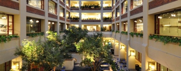
Interior photo of MGH’s Building 149 at the Charlestown Navy Yard, where much of our research on healthy older adults is conducted.

Beyond the lab itself, there are other major differences between my career and that of the typical academic. Another big, big difference is that because there are no students, there is no teaching (and alas, no summer breaks)! Many academics in the university model must negotiate and balance teaching alongside of research. Here at HMS, tenure and promotion is based purely on clinical work, research productivity, and hospital / community service. Less-formal and informal teaching opportunities are still abundant, however. For example, I have convened two statistical courses this year within my lab, one for the research assistants and one for the doctoral researchers. And through the Harvard Catalyst system, it is also possible to take or teach courses on an enormous variety of subjects, including career-oriented options like lab management and budgeting, advanced grantwriting, and research development.

The tenure track at HMS is also somewhat different from the university model. There is an added level below the assistant professor rank, called Instructor. Postdoctoral fellows tend to apply to the Instructor rank when they intend to remain with the lab they work in as a permanent option, rather than staying postdocs forever. Instructors tend to have much more stability than postdocs, receive higher salaries, and have access to a broader suite of resources at Harvard and MGH.

The path to tenure has also been tweaked. Although the main Harvard University campus was once notorious for making the path to tenure extremely difficult, there are very clear, achievable guidelines for the process at HMS. There is no tenure clock, meaning you can stay at the Instructor rank – or any other – as long as you’d like before applying to the next-higher level. This comes with a clear risk over typical university positions, though: The downside is that with no clock, there is no guaranteed salary support. This is often called a “soft money” position, and means that your salary comes exclusively through the grants you write and receive. This can be stressful when the funding climate becomes very competitive, but both HMS and MGH have several internal grant awards designed for funding gaps. So the reality is that although the money is soft, many researchers can go 4-5 years without a successful external grant before salary support becomes a problem.

All in all, I am very glad to be developing my career in a hospital setting, and particularly at HMS / MGH. This is because my job has far more flexibility than what I’d be doing in a university setting. Research can and must remain a primary focus, but beyond that, it’s really up to me. I can teach or not, I can see patients or not, I can set up my own mini-lab within my research group or not. I can take on some of these things temporarily and then give them up later without major consequences. Some people work within my research group indefinitely, while others enter the job market after a few years. While there’s a strong incentive to write and receive grants, HMS / MGH have developed a strong support structure to make it easier to succeed in grantwriting. This is supplemented by the support structure within my lab for grants and high-impact research.

cart-team

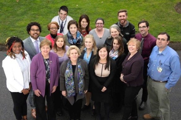
We wear many hats in my hospital laboratory. Here is the team that runs the Center for Alzheimer Research and Treatment, our clinical trials arm located at Brigham & Women’s Hospital.

All this adds up to the best part of my career: work-life balance. We definitely work hard, but it’s possible to take many weekends and evenings off to spend with family and loved ones. While the exact number of hours per week varies by personality, life stage, and whether a grant is coming due, nearly everyone in my lab has been able to find their optimal work schedule. I believe that this contributes to the strong diversity in my lab, which is made up of many parents and is about 70% female, including at the most senior levels of our group.

Things at HMS / MGH aren’t perfect, of course, but for those who wish for research options outside of the traditional lab-and-university environment, it’s a great “alternative” career with only a few tradeoffs from the standard academic model. Please leave questions and comments below – I’d be happy to expand on the details of my day-to-day.